
Myths vs. Facts: Dental X-Rays for Children
September 28, 2026
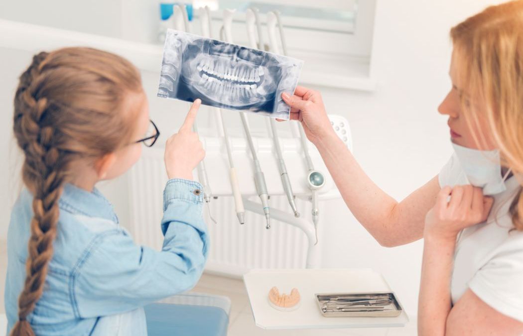


Dental X-rays are one of the most consistently questioned aspects of pediatric dental care. Parents who are thoughtful about their children's health often ask about radiation exposure, about frequency, and about whether X-rays are really necessary when no symptoms are present. These are reasonable questions — and they deserve accurate answers rather than dismissal.
At Shaenfield Pediatric Dentistry & Orthodontics in northwest San Antonio, the bilingual team provides these answers as a standard part of patient communication — because informed parents make better decisions about their children's care, and the questions about X-rays are both legitimate and answerable.
Myth: Dental X-Rays Expose Children to Dangerous Levels of Radiation
Fact: Digital pediatric dental X-rays deliver radiation exposure that is minuscule by any meaningful comparison.
Modern digital X-ray technology has dramatically reduced radiation exposure compared to the film X-rays of previous decades. A standard set of bitewing X-rays delivers approximately 0.005 millisieverts (mSv) of radiation — for context, a cross-country flight delivers approximately 0.03 mSv, and natural background radiation from everyday environmental sources delivers approximately 3 mSv per year.
The radiation from a pediatric dental X-ray is a fraction of a percent of what a child is exposed to from natural background sources annually. The size-adjusted collimators and digital sensors used at Shaenfield Pediatric Dentistry minimize exposure specifically for smaller patients. The concern about radiation from dental X-rays is understandable given the general cultural anxiety around radiation — but the actual numbers don't support it.
Myth: X-Rays Aren't Necessary If Nothing Hurts and Nothing Looks Wrong
Fact: The conditions dental X-rays most reliably catch are asymptomatic — which is precisely why clinical examination alone isn't sufficient.
Interproximal decay (cavities between teeth) is invisible on clinical examination. A dentist looking directly at a child's teeth cannot see the early-stage cavity developing between two molars at the contact point. The first sign of that cavity, without radiographic evaluation, may be the child reporting pain — by which point the decay has progressed significantly.
The same applies to bone levels (which identify developing gum disease), early pathology (including cysts and developing tumors that are asymptomatic until significantly advanced), and the position and development of unerupted permanent teeth. These findings are radiographic — they exist below the surface and are invisible to visual examination.
A "nothing looks wrong clinically" assessment and a "nothing is developing radiographically" assessment are fundamentally different. Pediatric dentistry without radiographic evaluation is dentistry with a significant diagnostic blind spot.
Myth: Children Need X-Rays at Every Single Appointment
Fact: X-ray frequency is determined by individual risk factors, not a one-size-fits-all schedule.
The American Dental Association and American Academy of Pediatric Dentistry guidelines for X-ray frequency are risk-stratified. Children with a history of cavities, dietary patterns high in sugar, poor plaque control, or crowded dentition that makes clinical examination difficult are candidates for more frequent radiographic evaluation — typically every six to twelve months for bitewings. Children at lower risk with no history of decay and adequate home hygiene may appropriately have radiographs at less frequent intervals.
At Shaenfield Pediatric Dentistry & Orthodontics, the decision about when to take X-rays is made based on each child's specific risk level and clinical picture — not a default protocol applied regardless of circumstances.
What Pediatric X-Rays Actually Show
The diagnostic value of routine pediatric dental X-rays is specific and meaningful:
- Interproximal decay: The cavities between teeth that clinical examination cannot detect.
- Bone levels: Early changes in the bone supporting the teeth that indicate developing periodontal disease — not common in children but not absent either, particularly in children with specific systemic conditions.
- Developing tooth buds: The position, development, and eruption path of unerupted permanent teeth. Panoramic X-rays in particular reveal whether permanent teeth are developing normally, whether any are missing (hypodontia), whether any are erupting in incorrect positions, or whether supernumerary (extra) teeth are present.
- Pathology: Cysts, tumors, and other pathological conditions that are often entirely asymptomatic in early stages and detectable only radiographically.
For the combined pediatric dental and orthodontic care Shaenfield provides, the radiographic record that tracks developing dentition, eruption sequence, and jaw growth is an essential diagnostic tool — not an optional one.
Schedule Your Child's Appointment at Shaenfield Pediatric Dentistry & Orthodontics
The bilingual team at Shaenfield Pediatric Dentistry & Orthodontics serves families in New Territories, Northwest Crossings, Saddlebrook Farms, Oak Grove, Braun Station, Wildwood West, and surrounding northwest San Antonio communities at 11590 Galm Road, Suite 105. Flexible scheduling and family block appointments make getting everyone in practical. Call (210) 672-4200 to schedule. The questions about X-rays are good ones — and the answers make the decision clearer.
How Often Should Children Have Dental X-Rays?
For children at average cavity risk with no history of decay and good home hygiene, bitewing X-rays are typically appropriate every twelve to twenty-four months. For children at higher risk — with prior cavities, dietary patterns heavy in sugar, or compliance challenges with brushing — a six-to-twelve month interval is more appropriate.
Panoramic X-rays that show the full developing dentition and jaw architecture are typically taken at specific developmental points — the early mixed dentition period around age 6 to 7, and again in the early teen years when comprehensive orthodontic planning benefits from a full view of tooth development and eruption sequence.
At Shaenfield Pediatric Dentistry & Orthodontics, the combined pediatric dental and orthodontic care model means that the radiographic record serves both purposes — tracking decay risk and tracking developing dentition for orthodontic planning — without unnecessary duplication.
Bilingual Care for Northwest San Antonio Families
The bilingual team at Shaenfield ensures that every conversation about X-rays, dental care, and orthodontic treatment happens in the language that is most comfortable and most clearly understood. For families in New Territories, Northwest Crossings, Saddlebrook Farms, and the surrounding northwest San Antonio communities, accessible communication is part of what makes the practice a trusted choice across generations of families.
The exam that finds something in the X-ray today is always better than the emergency appointment that follows the problem those X-rays could have identified months or years earlier.


